Our oxygen probes are based on metalloporphyrins, in which both the formation of triplet states via intersystem crossing (S1→T1) and the subsequent phosphorescence occur with high quantum efficiencies. To ensure biological compatibility and selectivity for oxygen, porphyrins are encapsulated within hydrophobic dendrimers, whose periphery is modified with polyethyleneglycol (PEG) residues [3, 4, 5 ]. The resulting constructs are inert, hydrophilic and fully bio-compatible. The phosphorescence lifetimes of these probes provide a selective signal for oxygen and allow measurements of absolute oxygen concentrations in any aqueous environment. Measurements based on phosphorescence lifetimes are independent of the probes' concentrations and unaffected by the optical properties of the medium.
Recently, the capabilities of the method have been extended by combining it with two-photon laser scanning microscopy. The resulting technique is known as two-photon phosphorescence lifetime microscopy (2PLM) of oxygen [6]. 2PLM makes it possible to assess tissue oxygen metabolism with high spatial resolution at depth in such areas as the brain cortex [7, 8], the retina of the eye [9,10], bone marrow [11, 12, 13] and newly growing bone [14], thus addressing biological questions for which non-invasive imaging of oxygen provides critical information not obtainable by any other method. More clinically relevant applications include measurements in the intestines [15, 16] and in tumors undergoing radiation therapy [17].
One major advantage of phosphorescence is its long lifetime, which allows for complete suppression of scattered excitation and tissue autofluorescence using microsecond time-gating. We are developing phosphorescent probes for quantitative imaging of temperature, pH and metal ions simultaneously with oxygen. These probes are constructed around multichromophoric systems, in which energy and electron transfer processes are tuned to render desired quantitative analyte-specific signals.

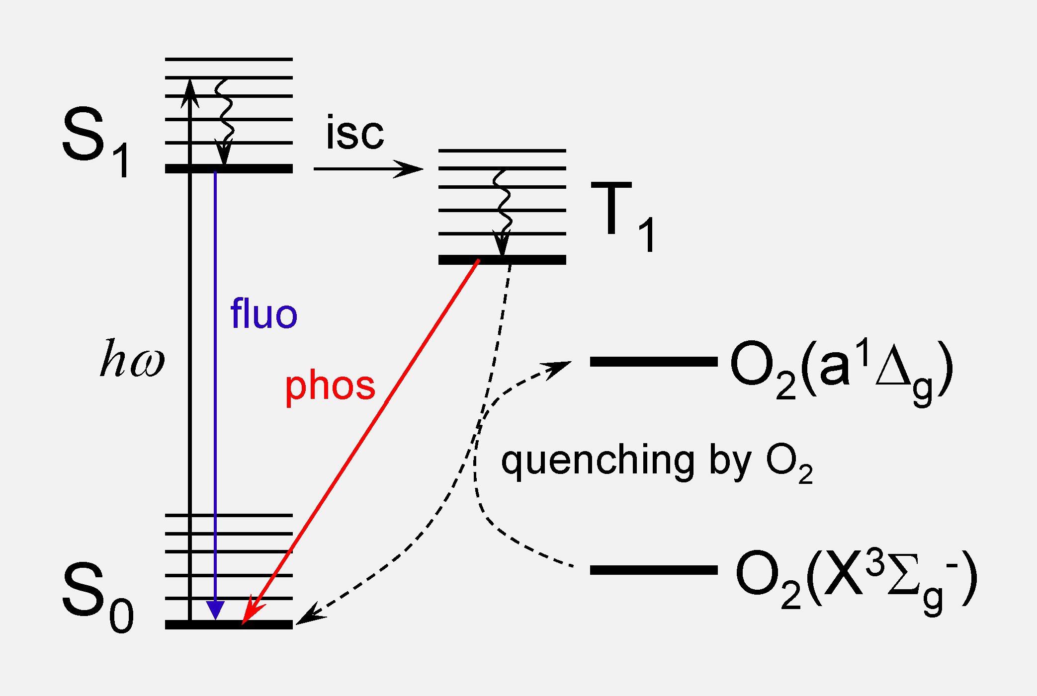 Energy diagram illustrating basic photophysical processes in a probe molecule and quenching of the triplet state by oxygen.
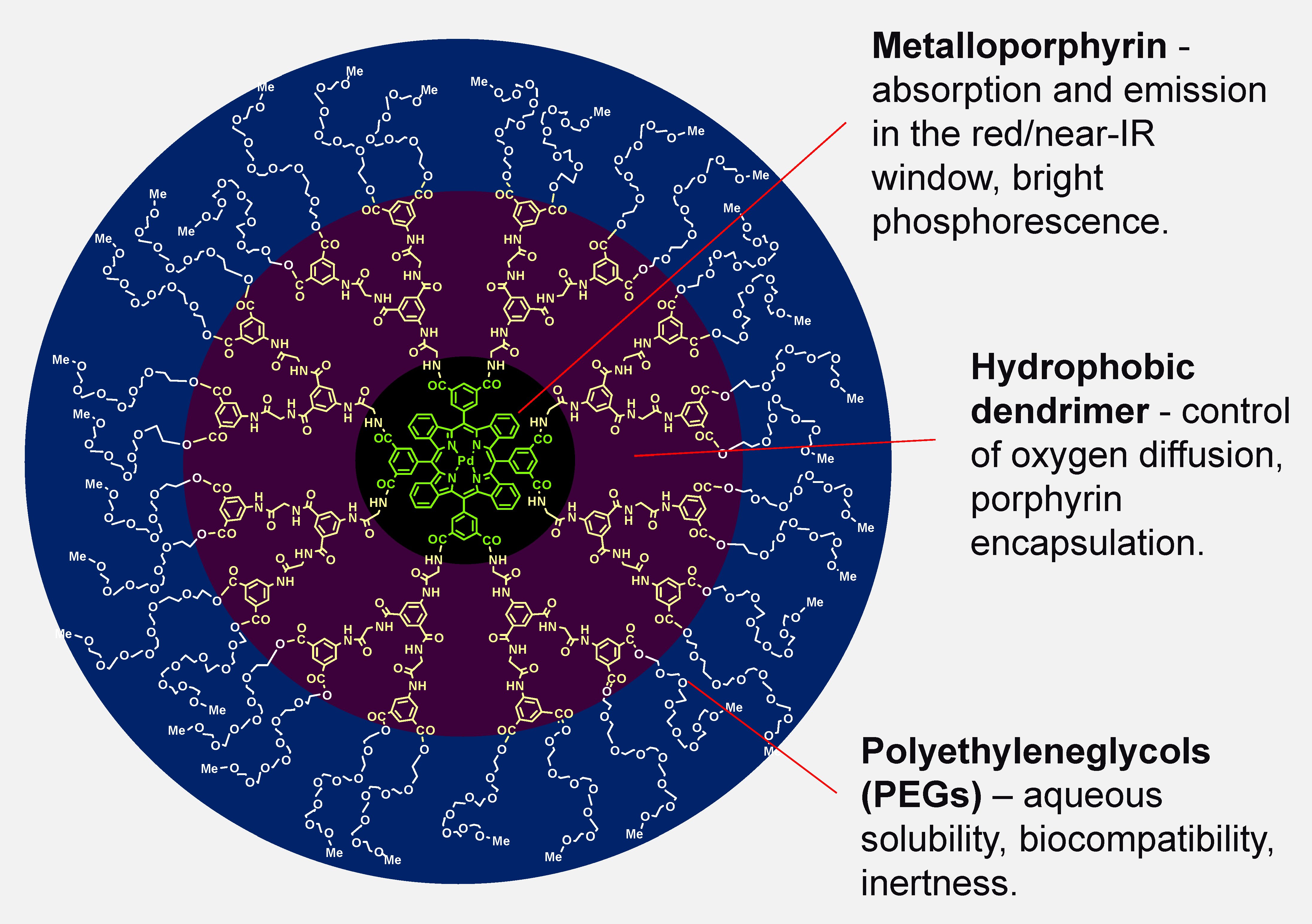 Structure of a dendritically protected phosphorescent probe for O2.
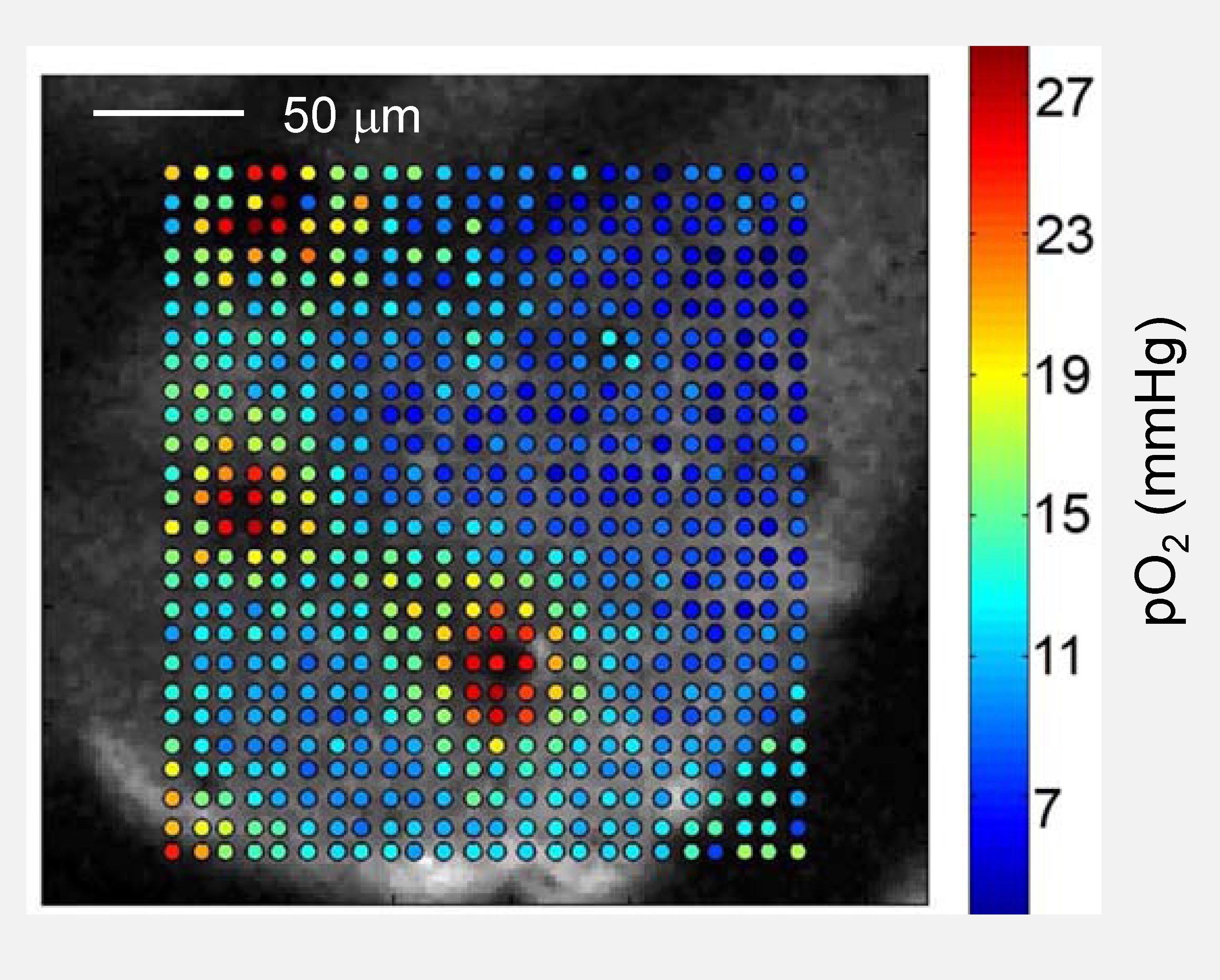 High-resolution oxygen map in the mouse brain obtained using 2PLM. Depth: 80 μm.
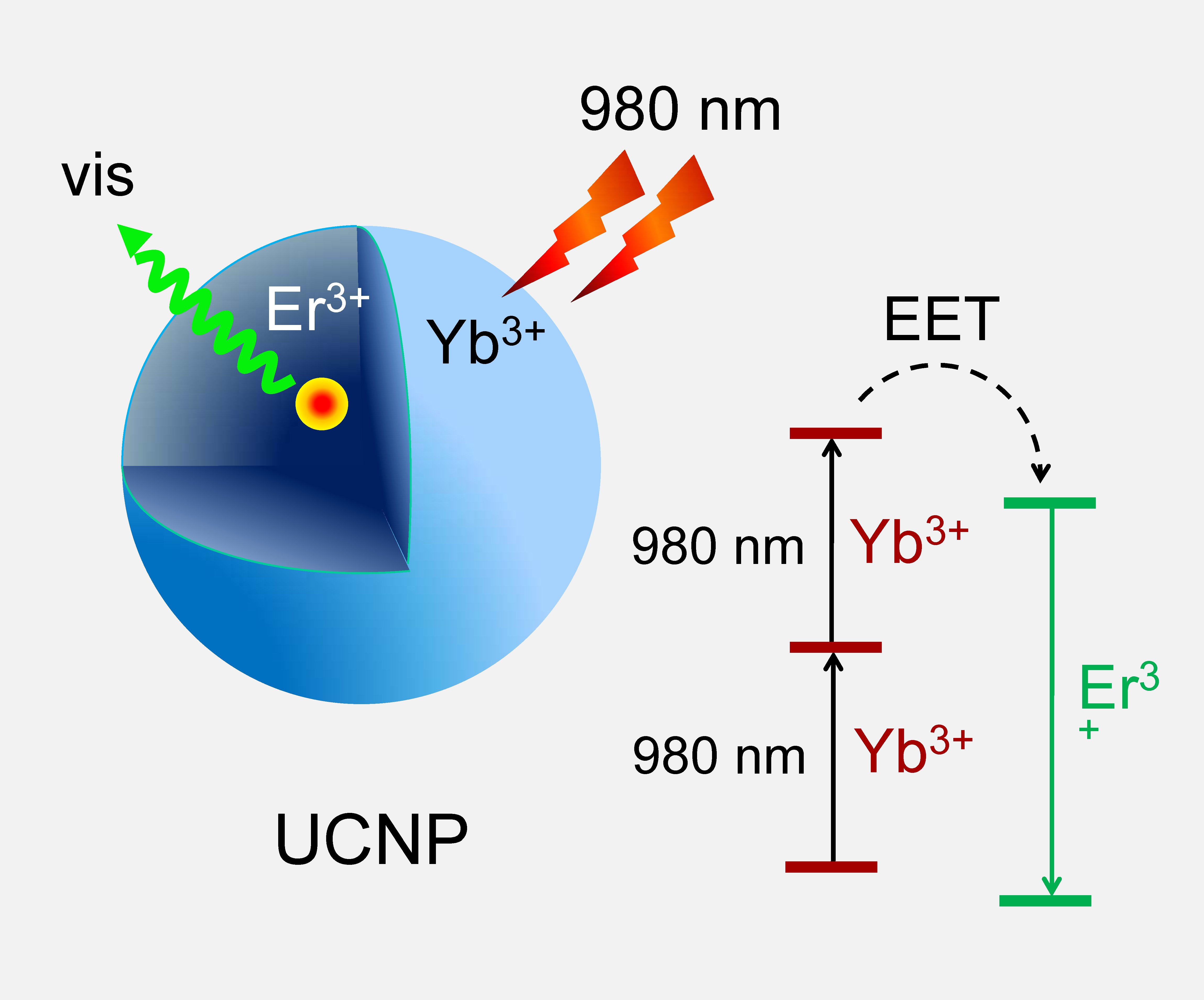 Sequential two-photon excitation and energy upconversion in UCNP crystals.
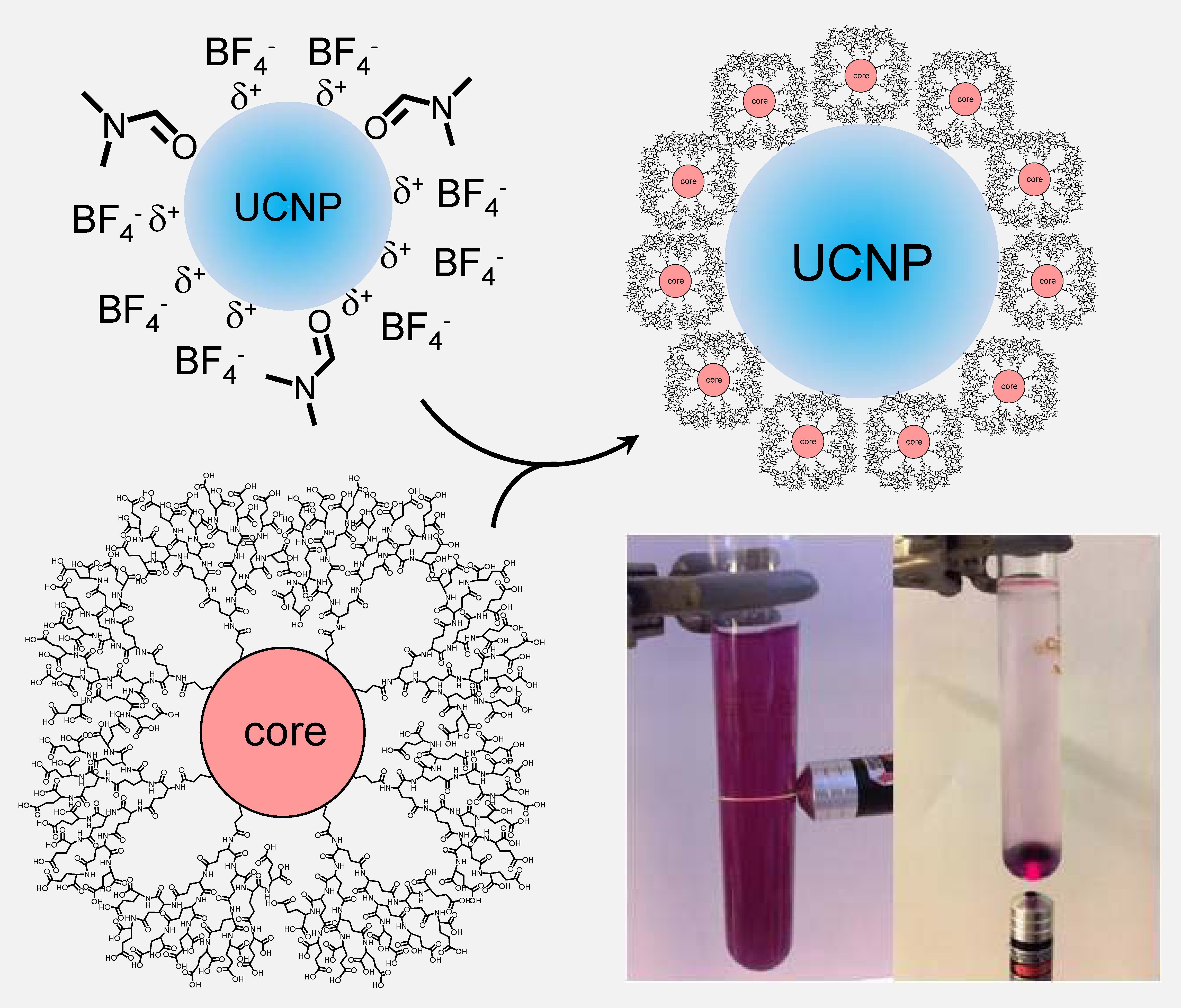 Solubilization of UCNPs using hydrophilic dendrimers. Upconverted emission from an aqueous solution of dendritic UCNPs excited by a hand-held laser pointer (980 nm).
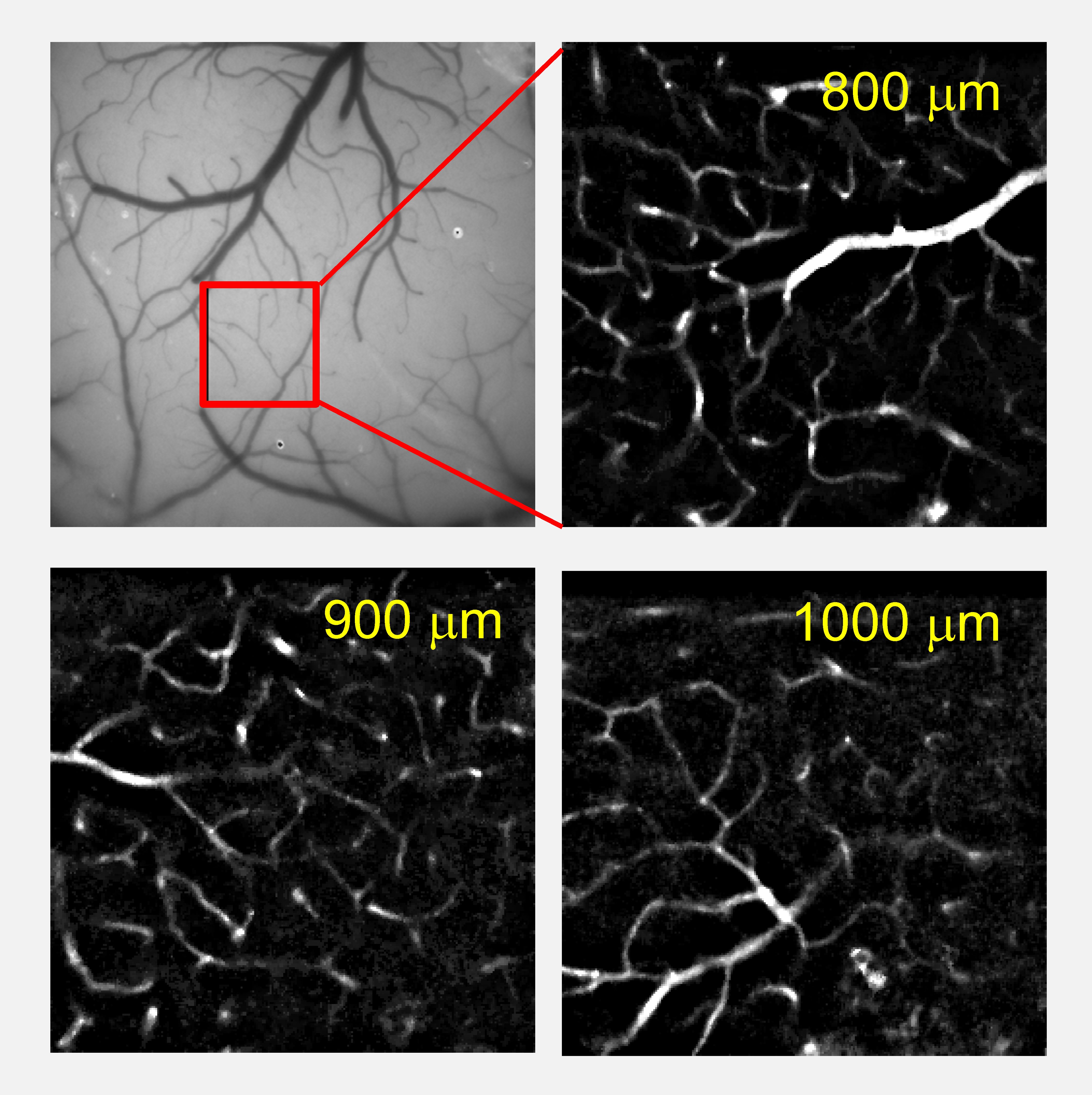 High-resolution depth-resolved maps of brain vasculature in mice obtained using dendritic UCNPs and two-photon CW excitation with average power not exceeding 20 mW.